Early detection - Effective arsenal in the fight against cancer
The development and implementation of cost-effective screening methods for detecting cancer early in its course when it is most responsive to treatment is one of the most direct ways to address the health disparities in tobacco-related cancer. Several “liquid biopsy” techniques are being developed in California and hold the most promise for low-cost, easy-to-use early detection methods that can be administered at a variety of community clinics throughout the state.
Cancer is one of the leading causes of death in California, second only to heart disease. One out of 10 Californians were diagnosed with lung or colorectal cancers in 2012, both of which are directly related to tobacco use. Even though the incidence of other cancers is higher, with approximately three out of 10 Californians diagnosed with either breast or prostate cancer, lung cancer remains the leading cause of cancer death in California. This is particularly true among African-American men and women who both die from lung cancer at a disproportionally higher rate than their white, Hispanic, and Asian/Pacific Islander counterparts.
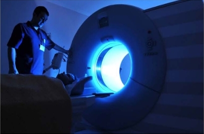
The development and implementation of cost-effective screening methods for detecting cancer early in its course when it is most responsive to treatment is one of the most direct ways to address the health disparities in tobacco-related cancers. The recent approval of Medicare coverage of screening for lung cancer with low dose computed tomography (LDCT) demonstrates the importance that the healthcare community places on early detection. Although a step in the right direction, the average cost of an LDCT scan is $300 and can vary widely among different clinics, making it less accessible to low-income patients – one of the primary populations that have a disproportionately high lung cancer incidence. Beyond LDCT, tremendous advances have recently been made in the development of low-cost accurate methods for detecting cancer early using a patient’s blood or even saliva. Several of these “liquid biopsy” techniques are being developed in California and hold the most promise for low-cost, easy-to-use early detection methods that can be administered at a variety of community clinics throughout the state.
Below are some of the TRDRP-funded research findings related to cancer early detection:
A key strategy to early detection methods is the identification and characterization of “biomarkers”, i.e., molecules that are found in abundance in cancerous tissues but are not readily found in non-diseased tissues. The molecules may be proteins, genetic molecules called nucleotides, or even sugars. Very often these molecular differences appear very early in the course of the disease, even before a doctor would classify it as definitive cancer. The molecules, or the cells they are attached to, can shed from the original site, e.g., a pre-cancerous tumor in the lung, and enter the blood circulation of the patient. By counting the number of these cancer biomarker molecules in the blood, a doctor can make a prediction as to whether the patient is likely to develop cancer, even if it is not visible on chest x-ray or LDCT.
The report by Clarke et al. describes their study of a protein that is being investigated as a potential a biomarker for lung cancer. The researchers are studying the function of the protein to determine whether it may also give a clue as to how it causes cancer and whether a therapy can be developed to prevent that progression to cancer.
Clarke N. et al., PLoS ONE 2015, 10(11): e0142061
The ideal biomarker would be a molecule that is only made by cancer cells and is never made by any normal cell in the body. Therefore, if even one such molecule was detected in a patient then there would be no doubt that the patient has cancer. Such a biomarker is said to have “high specificity” for cancer or has a “low false-positive” rate. In reality, most biomarkers are molecules that are made by both cancer and normal cells and it is the amount that is made by tumor cells that is the main difference. New technological developments have enabled researchers to identify multiple biomarkers for a single cancer type which increases the specificity of the detection method. Below are studies that are developing such multiplexed biomarker detection methods using 32 different proteins to detect oral cancer (Xiao et al.), 29 different sugars (Ruhaak et al.) and 12 genetic molecules called “nucleotides” (Zhang et al.) to detect lung cancer .
Xiao et al., Oral Oncology 2015, 51, 1011–1019
Ruhaak et al., J. Proteome Res. 2015, 14, 4538−4549
Zhang et al., Cell. Mol. Life Sci. 2012, 69:3341–3350
Annual screening of high-risk patients with low-dose computed tomography (LDCT) was approved for Medicare coverage in February 2015. These scans are very similar to regular CT or CAT scans except that they expose the patient to a much lower dose of radiation, minimizing the chance of adverse radiation effects occurring in patients that are screened on a yearly basis. One concern of LDCT is that it produces poorer quality images than regular CT and may prevent a doctor from seeing a tumor (i.e., false-negative) or cause a doctor to misdiagnose a normal lung structure as cancer (i.e., false-positive). The report by Young et al. tests the agreement of diagnoses from different doctors interpreting the same LDCT scans and suggests enhancements to the procedure that can improve the agreement. The report by Chong et al. describes a computer program that doctors can use to help distinguish between cancerous and non-cancerous lesions on LDCT scans, thereby reducing the number of false-positive and false-negative interpretations.